
Our Office
Mansfield Family Dentistry
100 Carlin Road
Mansfield, TX 76063
Phone: (817) 473-0291
Fax: 682-518-1190
Visit Us Online

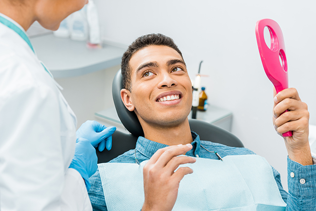
At the office of Mansfield Family Dentistry, we take a practical, prevention-first approach to oral health. Regular oral exams are the cornerstone of that approach — they give our team a clear snapshot of your current dental status, let us catch problems while they're small, and help patients of every age keep their smiles healthy and functional. During each visit we combine careful examination with education so you leave with a clear understanding of your oral health and the next steps, if any, that are recommended.
Your initial exam sets an essential baseline. We'll begin by reviewing your medical and dental histories, paying special attention to medications, previous dental work, and any symptoms or concerns you bring. Sharing details about your habits — from grinding or clenching to tobacco use — helps us tailor the exam and any recommendations that follow.
The clinical portion of the visit is systematic and thorough. Our dentists and hygienists examine each tooth, evaluate gum health, check bite relationships, and assess TMJ (temporomandibular joint) function. If needed, we take targeted diagnostic images to evaluate the roots, bone, and structures that are not visible during a visual inspection. A periodontal screening is included when appropriate to measure how well the supporting tissues are holding up.
Communication is a priority. After the exam and review of any radiographs, we discuss findings in plain language and outline options — from homecare adjustments to monitoring or intervention. We take time to answer questions and make sure you understand the purpose of any suggested care so you can make informed decisions about your dental health.

Oral exams do more than protect teeth and gums; they can reveal signs of broader health issues. Many systemic conditions show early clues in the mouth — from persistent dry mouth and unusual ulcerations to gum changes or persistent bad breath. Detecting these signs early helps patients seek medical evaluation when appropriate.
Research continues to show links between oral inflammation and systemic disease. During an exam we look for patterns that may suggest increased risk for other health problems and discuss when a conversation with your physician might be warranted. Keeping the mouth healthy is an important part of overall well-being.
Below are some conditions that are commonly associated with oral health issues and that our clinicians remain vigilant for during routine exams:
Digestive disorders
Respiratory illnesses
Cardiovascular disease
Stroke
Diabetes
Neurological conditions, including links being explored with Alzheimer's disease
Likewise, some medical treatments and conditions can produce oral symptoms. Our exam process is designed to recognize changes that suggest a systemic origin so we can coordinate care with your other healthcare providers when necessary.

Daily brushing and flossing form the foundation of good oral health, but professional checkups and cleanings fill the gaps that homecare can miss. A hygienist removes hardened deposits and soft plaque from areas that are difficult to access, polishing away surface stains and leaving the mouth feeling refreshed.
Regular visits also let us monitor small changes over time. By checking teeth, restorations, gum levels, and bite dynamics at scheduled intervals, we can spot early decay, beginning bone loss, or wear patterns caused by grinding. Early detection generally leads to simpler, more conservative treatment options.
These appointments are also an opportunity for personalized coaching. We demonstrate effective brushing and interdental cleaning techniques, review diet and habits that influence oral health, and recommend preventative treatments (such as sealants or desensitizing therapies) for patients who can benefit from them.
Children and adolescents benefit from consistent monitoring of growth and development. Routine exams help us determine when orthodontic intervention might be helpful and allow us to guide families on establishing healthy habits that protect teeth throughout life.
Visual inspection tells us a lot, but diagnostic images reveal what lies beneath the surface. Dental radiographs help identify decay between teeth, evaluate root structure, check bone health, and detect pathology that can't be seen with the naked eye. They are an essential part of a comprehensive oral exam when indicated.
Digital radiography has become the standard because it provides immediate images with very low radiation exposure. Images are captured quickly, reviewed on-screen with you, and securely stored in the patient file to track changes over time. This technology supports clearer communication and faster decision-making.
Digital radiography offers several advantages to patients and clinicians, including:
Reduced radiation exposure compared with traditional film methods
Immediate viewing so findings can be discussed during the same visit
Ease of sharing and collaboration with specialists when a referral is needed
Environmentally friendly workflow with no chemical development processes
Secure, long-term storage in the patient's electronic record for monitoring changes
The images we select depend on the clinical question. Different techniques highlight different structures and are used for specific diagnostic or treatment-planning purposes. Below are some of the most common types and why they might be recommended.
Periapical x-ray - Focuses on a single tooth from crown to root and is useful for diagnosing root problems or localized infection.
Bitewing x-ray - Shows the upper and lower back teeth in one view and is particularly effective for detecting interproximal decay and evaluating restoration margins.
Full Mouth Series - A comprehensive collection of periapical and bitewing images that offers a detailed overview of all teeth and surrounding bone.
Panoramic Film (panorex) - A broad, two-dimensional view of the jaws, teeth, and surrounding structures used for screening growth, development, impacted teeth, and jaw pathology.
Cephalometric Film - A side-profile image used primarily in orthodontics to evaluate skeletal relationships and treatment planning.
For more complex diagnostic needs, three-dimensional imaging such as cone-beam computed tomography (CBCT) can be employed. CBCT provides a volumetric view of teeth, bone, and surrounding anatomy and is particularly helpful for surgical planning and precise implant placement when applicable.
At every step, our goal is to use the least-invasive, most informative tools available to produce a clear diagnosis and a patient-centered plan of care. When imaging is recommended, we explain the reason, the process, and how it will inform treatment decisions.
In summary, routine oral exams are a powerful tool for protecting both oral and overall health. They combine careful clinical assessment, targeted imaging, and patient education to help prevent disease and preserve function. If you have questions about what to expect at an exam or would like more information, please contact us to learn more.
An oral exam is a comprehensive clinical assessment of the teeth, gums, soft tissues, jaw joints and bite that helps identify current problems and risk factors for future disease. It is the foundation of preventative dental care because it allows clinicians to detect early signs of decay, gum disease, oral cancer and functional issues before they become more complex. A thorough exam combines visual inspection, tactile assessment and targeted diagnostic imaging when indicated to produce a clear clinical picture.
Regular oral exams support long-term oral function and comfort by guiding personalized homecare and professional maintenance plans. When exams are performed consistently, changes can be tracked over time so treatment is more conservative and outcomes are better. At Mansfield Family Dentistry we emphasize education and shared decision-making so patients understand findings and next steps.
Your first oral exam establishes a baseline and begins with a review of medical and dental history, medications and any concerns you bring to the visit. The clinician will ask about habits that influence oral health such as tobacco use, grinding or dry mouth to tailor the assessment and recommendations. Understanding your history helps us interpret clinical findings and plan appropriate preventative or restorative care.
The clinical portion includes a systematic check of each tooth, an evaluation of gum health and a review of bite relationships and TMJ function. When needed, we take targeted digital images to evaluate areas not visible during the visual exam and perform a periodontal screening to measure supporting tissue health. The visit concludes with a clear explanation of findings, suggested next steps and time for questions so you leave informed and comfortable with the plan.
Frequency of oral exams depends on individual risk factors such as personal medical history, gum health, past decay, restorative work and lifestyle habits. For many patients a six-month interval is appropriate because it balances preventative care with ongoing monitoring, but some people may benefit from more frequent visits while others can safely extend intervals. Your clinician will recommend an exam schedule tailored to your needs based on findings and risk assessment.
Regularly scheduled exams allow clinicians to detect small changes early, which often leads to simpler, less invasive treatment and better long-term outcomes. Maintaining a consistent exam cadence supports tracking of restorations, bone levels and wear patterns and helps identify when additional interventions or specialist referrals are needed. The exam schedule can be adjusted over time as your oral health status evolves.
The mouth often reflects broader health trends, and changes observed during an oral exam can signal systemic issues such as diabetes, nutritional deficiencies or immune disorders. Clinicians look for persistent dry mouth, unusual ulcerations, bleeding gums or mucosal changes that may warrant medical evaluation. Early recognition of these oral signs can prompt timely communication with your physician and support comprehensive care.
Research continues to reveal links between oral inflammation and conditions like cardiovascular disease and metabolic disorders, so periodontal screening is an important component of a whole-body health approach. When patterns suggest elevated systemic risk, clinicians will document findings and suggest appropriate follow-up with medical providers. Coordinated care helps ensure both oral and overall health are managed effectively.
Diagnostic imaging complements the visual exam by revealing structures beneath the surface such as tooth roots, bone levels and hidden decay. Common modalities include periapical images for single-tooth evaluation, bitewings to check for interproximal decay and restoration margins, and panoramic films for a broad overview of the jaws, impacted teeth and developmental concerns. Each image type answers a specific clinical question and is selected for its diagnostic value.
Digital radiography has become standard because it produces immediate images with very low radiation exposure and allows the team to review findings with patients during the same visit. For complex evaluations, three-dimensional imaging such as cone-beam computed tomography (CBCT) may be used to visualize bone anatomy and spatial relationships for treatment planning. When imaging is recommended, clinicians explain the purpose, the process and how the information will influence care decisions.
Images are stored securely in the patient record to support longitudinal monitoring and to facilitate referrals when specialist collaboration is needed. The goal is always to use the least-invasive, most informative tools necessary to reach a confident diagnosis and to guide conservative, patient-centered treatment planning.
Modern dental imaging uses very low radiation doses and adheres to strict safety protocols to minimize exposure while providing essential diagnostic information. Digital radiography reduces radiation compared with traditional film and delivers images instantly, which shortens time in the operatory and improves workflow. Protective techniques and equipment, such as lead aprons and thyroid collars when appropriate, further reduce risk for patients.
Clinicians follow the principle of ALARA (as low as reasonably achievable) when ordering images, balancing diagnostic benefit against exposure. Imaging is recommended only when it will change diagnosis or treatment planning, and frequency is individualized based on clinical need and patient risk factors. If you have concerns about radiation, your clinician can discuss the specific benefits and safety measures related to the recommended images.
Oral cancer screening is an integral part of a comprehensive oral exam and involves a careful inspection and palpation of the lips, oral mucosa, tongue, floor of mouth and oropharynx to identify any suspicious lesions or lumps. Clinicians look for persistent sores, white or red patches, unexplained lumps or areas that bleed easily, and they document any findings for ongoing monitoring. Early detection significantly improves treatment options and outcomes, so routine screening is strongly emphasized.
Adjunctive tools such as tissue fluorescence or VELscope screening may be used to enhance visualization of abnormal tissue in select cases, but these tools do not replace a thorough clinical exam. When a lesion of concern is identified, clinicians will recommend appropriate follow-up which may include short-term monitoring, referral to an oral medicine specialist or biopsy for definitive diagnosis. Clear communication about findings and next steps is provided so patients understand the process and the reasons for additional evaluation.
Professional cleanings performed by a dental hygienist remove hardened tartar and biofilm from areas that are difficult to reach with routine homecare, which helps reduce inflammation and lower the risk of periodontal disease. Cleanings also polish the teeth, remove surface stains and leave the mouth feeling refreshed while allowing the clinician to assess the response of gum tissues to therapy. The combination of clinical exam and professional hygiene enables more accurate assessment of oral health status.
During the appointment hygienists provide personalized coaching on brushing and interdental cleaning techniques and recommend preventative measures such as sealants or desensitizing therapies when indicated. Regular professional maintenance supports long-term stability of restorations, helps preserve supporting bone and can prevent the progression of early disease. The hygienist and dentist work together to ensure a coordinated plan that meets the patient’s needs.
Pediatric and adolescent exams focus on growth, development and the establishment of healthy habits while also monitoring for early signs of decay or orthodontic concerns. Clinicians assess eruption patterns, spacing, jaw development and habits such as thumb-sucking or prolonged pacifier use that can affect alignment. Age-appropriate education for both the child and caregiver is provided to reinforce effective homecare and dietary choices that support long-term oral health.
Routine exams during childhood allow clinicians to intervene early when interceptive orthodontic treatment or preventative measures like sealants are beneficial. Monitoring spacing and growth helps determine optimal timing for orthodontic evaluation and can reduce the need for more invasive treatment later. The team aims to create a positive, informative experience that encourages lifelong oral health habits.
Prepare for your exam by bringing a current list of medications, any recent medical updates and details about dental concerns or symptoms you have noticed, such as sensitivity, pain or chewing changes. If you have previous dental records or imaging from another provider, bringing them can help the clinician compare findings and streamline diagnosis. Arriving with specific concerns or goals allows the team to tailor the visit to your needs and to address priorities efficiently.
Useful questions to ask include what the clinician found on the exam, whether any images are recommended and how findings affect your short- and long-term care plan. Ask about prevention strategies specific to your risk factors, homecare tips, and how often follow-up exams should be scheduled. Clear communication helps you make informed decisions and ensures the exam visit supports your overall health and oral function.

Dental Implants, Root Canals, Sedation Dentistry, Tooth Extractions, Porcelain Veneers, Crowns, Dentures, Bridges, Nitrous Oxide, Teeth Whitening, Inlays & Onlays, Bonding, Fillings & Restorations, Dental Sealants, Invisalign®, Sleep and Snoring Issues
Our mission is to transform smiles and lives through exceptional, personalized dental care.
Mansfield Family Dentistry serves the Texas communities of Mansfield TX, South Arlington TX, Watsonville TX, Kennedale TX, Sublett TX, Britton TX, Venus TX, Midlothian TX, Irving TX, Grand Prairie TX, Haltom City TX, Bedford TX, Burleson TX and the Greater Arlington TX Area


